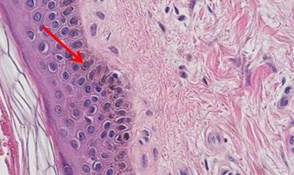
What makes the Hematoxylin and Eosin staining technique so essential in histology and pathology? This widely used technique highlights cellular structures, enabling detailed tissue analysis. Its simplicity and effectiveness make it a cornerstone in diagnostics and research. Let’s explore the step-by-step process for preparing the Hematoxylin and eosin (H&E) technique.
Preparing the Tissue Sample
The first step in H&E staining is preparing the tissue sample. Proper preparation ensures that the structural integrity of the tissue is maintained for accurate analysis. Tissues are first collected and fixed in a solution like formalin. Fixation preserves the cells and prevents degradation.
The sample is then embedded in paraffin wax to make it easier to slice into thin sections. These thin slices, usually 4-5 micrometers thick, are then placed on microscope slides. Proper preparation is critical for achieving consistent and clear staining results.
Staining the Tissue
Once the sample is prepared, the actual H&E technique begins. The technique involves two key steps: staining with hematoxylin and then with eosin.
Key steps in the staining process include:
- Deparaffinization:Removing the paraffin wax from tissue sections.
- Rehydration:Rehydrating tissues with decreasing concentrations of alcohol.
- Hematoxylin staining:Immersing tissues in hematoxylin to stain the nuclei.
- Rinsing:Washing excess dye to enhance clarity.
- Eosin staining:Applying eosin to stain cytoplasm and connective tissue.
- Dehydration:Removing water through increasing concentrations of alcohol.
- Mounting:Covering the stained section with a coverslip for preservation.
This process ensures that different cellular components are vividly highlighted, providing clear and detailed images under a microscope.
Visualizing Cellular Structures
The H&E technique provides excellent contrast between different cellular components. Hematoxylin stains nuclei a deep blue-purple, while eosin stains cytoplasmic and extracellular structures pink.
This contrast helps pathologists differentiate between normal and abnormal tissues. For instance, the technique is commonly used to identify cancerous cells, observe tissue architecture, and diagnose inflammatory conditions. The vivid staining achieved by this method simplifies the identification of structural anomalies, making it an indispensable tool in pathology.
Common Applications
This technique has a wide range of applications in both diagnostics and research. Its versatility makes it a go-to technique in histological studies.
- Cancer diagnosis:Identifying tumors and analyzing their structure.
- Inflammatory diseases:Detecting cellular responses in affected tissues.
- Developmental studies:Observing tissue growth and differentiation.
- Biopsy evaluations:Examining tissue samples for abnormalities.
- Toxicology research:Assessing the effects of substances on tissues.
These applications demonstrate the importance of mastering this technique. Understanding the process enables researchers and pathologists to conduct precise and meaningful analyses.
Tips for Optimal Results
Achieving the best results with H&E requires attention to detail and consistency. Proper handling of samples and adherence to protocols are crucial. Ensure that tissue sections are of uniform thickness to avoid uneven staining. Use high-quality reagents and maintain clean laboratory equipment. Regular calibration of staining times for hematoxylin and eosin improves consistency.
Partnering with a reputable biospecimen provider ensures access to high-quality samples, minimizing variability in results. Such providers follow strict standards for collection, preservation, and documentation, which enhances the reliability of staining outcomes. Working with trusted sources ensures that the integrity of the specimens is maintained, supporting accurate and reproducible results in research and diagnostics. By following these practices, the results are clearer and more reliable, supporting accurate interpretations.
One essential method in histology and pathology is the H&E staining. Its ability to highlight cellular structures with clarity makes it invaluable for diagnosing diseases and advancing scientific research. From preparing tissue samples to applying dyes, mastering the steps of staining ensures precise and reliable results. Its applications in diagnostics and research demonstrate its importance in improving healthcare outcomes.


